Generic selectors
Exact matches only
Search in title
Search in content
Post Type Selectors

Description

Product Description

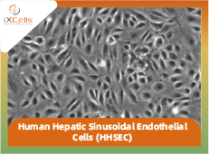
The human liver contains two distinct types of endothelial cells: vascular endothelial cells and sinusoidal endothelial cells. Human Hepatic Sinusoidal Endothelial Cells (HHSECs) are a specialized subset of microvascular endothelial cells that exhibit a distinct phenotype compared to other endothelial cell types. HHSECs function as antigen-presenting cells, facilitating antigen presentation to CD4+ T cells, and play a crucial role in regulating the immune response within the liver [1]. Additionally, HHSECs contribute actively to liver repair by serving as dynamic regulators that respond promptly and locally to zonal environmental stimuli [2].

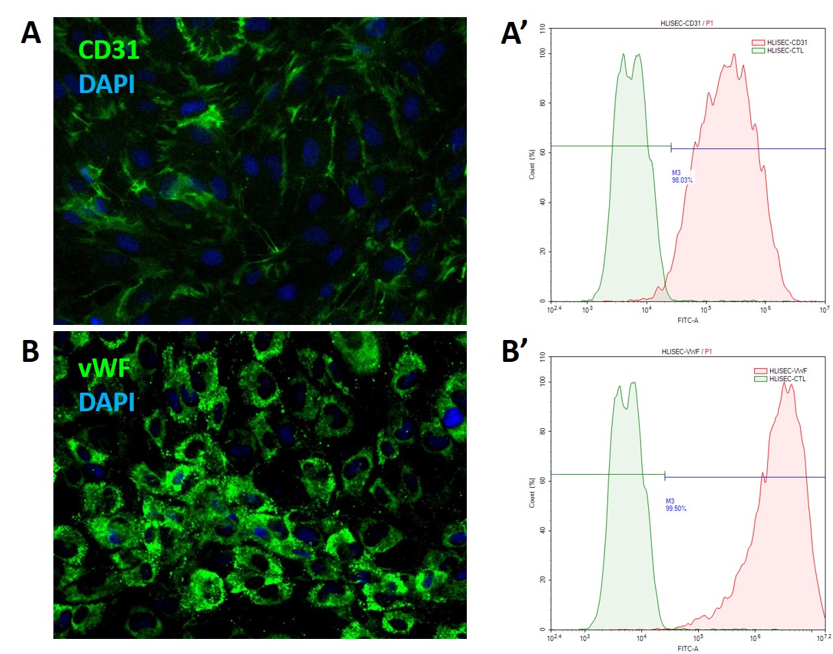
iXCells Biotechnologies provides high quality HHSECs, which are isolated from human liver and cryopreserved at P2, with ≥ 0.5 million cells in each vial. These HHSECs express CD31/PECAM-1, VE-Cadherin and von Willebrand Factor (vWF). They are negative for HIV-1, HBV, HCV, mycoplasma, bacteria, yeast, and fungi, and can be further expanded for no more than 3 passages under the conditions suggested by iXCells Biotechnologies. Further expansion may decrease the purity.

Figure 1.  Phase contrast images of Human Hepatic Sinusoidal Endothelial Cells (HHSEC, Adult). The cells were recovered and seeded at 10,000 cells/cm2 following iXCells’ protocol. The images were taken at the indicated time post-recovery.

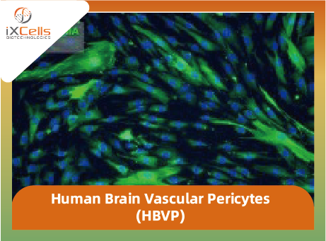
Figure 2. Immunofluorescence staining of HHSECs using antibodies against CD31/PECAM-1 (green), VE-Cadherin (red), and vWF (green). The nuclei were counterstained by DAPI (blue).

Product Details

Organism Homo Sapiens, Human
Cell Type Endothelial Cell
Tissue Human Liver
Disease Normal
Package Size 0.5 x 10cells/vial
Passage Number P2
Growth Properties Adherent
Product Format/Shipped Cryopreserved
Storage Liquid Nitrogen
Associated Media Endothelial Cell Growth Media

References

[1] Limmer A, and Knolle PA. Liver sinusoidal endothelial cells: a new type of organ-resident antigen-presenting cell. Arch Immunol Ther Exp (Warsz) 2001; 49 (1): S7-11.

[2] Wach KE etc. Sinusoidal ultrastructure evaluated during the revascularization of regenerating rat liver. Hepatoloty 2001; 33 (2): 363-378.

Datasheet & Culture Protocol

Technical Documentation

Citations

  • Jamil, M. A., Singer, H., Al-Rifai, R., Nüsgen, N., Rath, M., Strauss, S., . . . El-Maarri, O. (2020). Molecular analysis of fetal and adult primary human liver sinusoidal endothelial cells: A comparison to other endothelial cells. International Journal of Molecular Sciences, 21(20), 7776. doi:10.3390/ijms21207776 — Learn More

Product Highlights

      • The cells have been growing for an additional 2 passages upon recovery without antibiotics before release;
      • The endothelial cell-specific markers, including CD31, VE-Cadherin, and vWF, maintain >90% purity within 2-3 passages upon recovery.


Description

Product Description

Human liver contains two distinct endothelial cell types: vascular and sinusoidal. Human Hepatic Sinusoidal Endothelial Cells (HHSEC) are microvascular endothelial cells that exhibit a unique phenotype compared to other endothelial cells—they function as dendritic cells to present antigen for CD4+ T cells. Thus, HHSEC are actively involved in the regulation of the immune response in the liver [1]. HHSEC are actively involved in liver repair as dynamic regulators respond rapidly and locally to environmental zonal stimuli [2].

iXCells Biotechnologies provides high quality HHSEC, which are isolated from normal human liver and cryopreserved at P2, with >0.5 million cells in each vial. HHSEC express vWF/Factor VIII and CD31 (Figure 1). They are negative for HIV-1, HBV, HCV, mycoplasma, bacteria, yeast, and fungi and can further expand in Endothelial Cell Growth Medium (Cat# MD-0010) under the condition suggested by iXCells Biotechnologies.

Figure 1.  HHSECs provided by iXCells are positive for CD31 (A) and vWF (B), as shown by immunostaining. Flow analysis showed that more than 98% of cells are CD31 positive (A’), and more than 99% are vWF positive (B’).


Protocols
Thawing of Frozen Cells

1. Upon receipt of the frozen cells, it is recommended to thaw the cells and initiate the culture immediately in order to retain the highest cell viability.

2. To thaw the cells, put the vial in 37°C water bath with gentle agitation for 1-2 minutes. Keep the cap out of water to minimize the risk of contamination.

3. Prepare complete medium.

4. Pipette the cells into a 15 mL conical tube with 5 mL fresh Endothelial Cell Growth Medium (Cat# MD-0010).

5. Centrifuge at 1,000 rpm (~220 g) for 5 minutes under room temperature.

6. Remove the supernatant and resuspend the cells in fresh Endothelial Cell Growth Medium.

7. Culture the cell in T75 flask. Change the medium every other day until cells reach 80-90% confluence.

Safety Precaution: it is highly recommended that protective gloves and clothing should be used when handling frozen vials.


Standard Culture Procedure

1. HHSECs can be cultured in Endothelial Cell Growth Medium (Cat# MD-0010).

2. When cells reach ~80-90% confluence, remove the medium, and wash once with sterile PBS (5 mL/T75 flask).

3. Add ~2.5 mL of 0.25% Trypsin-EDTA to the flask and incubate for ~3 minutes at 37°C. Neutralize the enzyme by adding 2-3 volumes of cell culture medium.

4. Centrifuge 1,000 rpm (~220 g) for 5min and resuspend the cells in desired volume of medium.

8. Seed the cells in the new culture vessels at 5 × 10³ cells/cm2. Change the medium every other day until cells reach 80-90% confluence.


Product Details

  Tissue   Normal adult human liver
  Package Size   0.5 million cells/vial
  Passage Number   P2
  Shipped   Cryopreserved
  Storage   Liquid nitrogen
  Growth Properties   Adherent
  Media   Endothelial Cell Growth Medium (Cat# MD-0010)

References

[1]  Limmer A, and Knolle PA. Liver sinusoidal endothelial cells: a new type of organ-resident antigen-presenting cell. Arch Immunol Ther Exp (Warsz) 2001; 49 (1): S7-11.

[2]  Wach KE etc. Sinusoidal ultrastructure evaluated during the revascularization of regenerating rat liver. Hepatoloty 2001; 33 (2): 363-378.

Reviews

There are no reviews yet.

Be the first to review “Human Hepatic Sinusoidal Endothelial Cells (HHSEC)”

您的邮箱地址不会被公开。 必填项已用 * 标注

Related products