Generic selectors
Exact matches only
Search in title
Search in content
Post Type Selectors

Description

Product Description

Astrocytes are the major cell type in the mammalian brain. They provide a variety of supportive functions to their partner neurons in the central nervous system (CNS), such as neuronal guidance during development, and nutritional and metabolic support throughout life [1]. Astrocytes have also been implicated in various pathological processes [2] . Impairment of normal astrocyte functions during stroke and other insults can critically influence neuron survival. Long-term recovery after brain injury, through neurite outgrowth, synaptic plasticity, or neuron regeneration, is also influenced by astrocyte surface molecule expression and trophic factor release [3]. Numerous studies have demonstrated that astrocytes are among the most functionally diverse group of cells in the CNS. Much of what we have learned about astrocytes is from in vitro studies and astrocyte culture is a useful tool for exploring the diverse properties of this cell type.

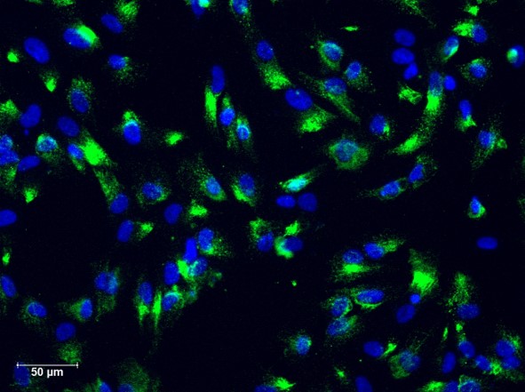
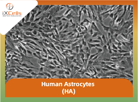
iXCells Biotechnologies provides high quality Human Astrocytes (HA), which are isolated from human brain (cerebral cortex) and cryopreserved at P2, with >0.5 million cells in each vial. These HA express GFAP (Figure 1). They are negative for HIV-1, HBV, HCV, mycoplasma, bacteria, yeast, and fungi. HA can further expand no more than 3 passages in Astrocyte Medium (Cat# MD-0039) under the condition suggested by iXCells Biotechnologies. Further expansion may decrease the purity and features of human astrocytes.


Human Astrocytes HAjpg

Figure 1. Human astrocytes (HA). (A) Phase contrast image of HA. (B) Immunofluorescence staining with antibody against GFAP.


Product Details

Tissue Human brain (cerebral cortex)
Package Size 0.5 millioncells/vial
Passage Number P2
Shipped Cryopreserved
Storage Liquid nitrogen
Growth Properties Adherent
Media Astrocyte Medium (Cat# MD-0039)

References

[1] G. I. Hatton (2002) Glial-neuronal interactions in the mammalian brain. Adv. in Physiol. Edu. 26:225-237.

[2] Van der Laan, L. J. W., De Groot, C. J. A., Elices, M. J. and Dijkstran, C. D. (1997) Extracellular matrix proteins expressed by human adult astrocytes in vivo and in vitro: an astrocyte surface protein containing the CS1 domain contributes to binding of lymphoblasts. J. Neurosci. Res. 50:539-548.

[3] Chen Y., and Swanson, R. A. (2003) Astrocytes and brain injury. J. Cereb. Blood Flow Metab. 23:137-149.


 

Download Datasheet

Technical Documentation

Citations

  • Yan, X., Jia, H., & Zhao, J. (2023). LncRNA MEG3 attenuates the malignancy of retinoblastoma cells through inactivating PI3K /AKT/mtor signaling pathway. Experimental Eye Research, 226, 109340. doi:10.1016/j.exer.2022.109340 — Learn More
  • Vidak, M., Jovcevska, I., Samec, N., Zottel, A., Liovic, M., Rozman, D., . . . Komel, R. (2018). Meta-Analysis and experimental VALIDATION Identified Frem2 and SPRY1 as NEW Glioblastoma Marker Candidates. International Journal of Molecular Sciences, 19(5), 1369. doi:10.3390/ijms19051369 — Learn More
  • Al-Serori, H., Ferk, F., Kundi, M., Bileck, A., Gerner, C., Mišík, M., . . . Knasmüller, S. (2018). Mobile phone specific electromagnetic fields induce transient dna damage and nucleotide excision repair in serum-deprived human glioblastoma cells. PLOS ONE, 13(4). doi:10.1371/journal.pone.0193677 — Learn More

Description

Product Description

Astrocytes are the major cell type in the mammalian brain. They provide a variety of supportive functions to their partner neurons in the central nervous system (CNS), such as neuronal guidance during development, and nutritional and metabolic support throughout life [1]. Astrocytes have also been implicated in various pathological processes [2]. Impairment of normal astrocyte functions during stroke and other insults can critically influence neuron survival. Long-term recovery after brain injury, through neurite outgrowth, synaptic plasticity, or neuron regeneration, is also influenced by astrocyte surface molecule expression and trophic factor release [3]. Numerous studies have demonstrated that astrocytes are among the most functionally diverse group of cells in the CNS. Much of what we have learned about astrocytes is from in vitro studies and astrocyte culture is a useful tool for exploring the diverse properties of this cell type.

iXCells Biotechnologies provides high quality Human Astrocytes (HA), which are isolated from human brain (cerebral cortex) and cryopreserved at P2, with >0.5 million cells in each vial. These HA express GFAP (Figure 1). They are negative for HIV-1, HBV, HCV, mycoplasma, bacteria, yeast, and fungi. HA can further expand in Astrocyte Medium (Cat# MD-0039) under the condition suggested by iXCells Biotechnologies.

Human Astrocyte

Figure 1. Immunofluorescence staining for GFAP

Product Details

  Tissue   Human brain (cerebral cortex)
  Package Size   0.5 million cells/vial
  Passage Number   P2
  Shipped   Cryopreserved
  Storage   Liquid nitrogen
  Growth Properties   Adherent
  Media   Astrocyte Medium (Cat# MD-0039)


References

[1] G. I. Hatton (2002) Glial-neuronal interactions in the mammalian brain. Adv. in Physiol. Edu. 26:225-237.
[2] Van der Laan, L. J. W., De Groot, C. J. A., Elices, M. J. and Dijkstran, C. D. (1997) Extracellular matrix proteins expressed by human adult astrocytes in vivo and in vitro: an astrocyte surface protein containing the CS1 domain contributes to binding of lymphoblasts. J. Neurosci. Res. 50:539-548.
[3] Chen Y., and Swanson, R. A. (2003) Astrocytes and brain injury. J. Cereb. Blood Flow Metab. 23:137-149.
Reviews

There are no reviews yet.

Be the first to review “Human Astrocytes (HA)”

您的邮箱地址不会被公开。 必填项已用 * 标注