
Description
Product Description
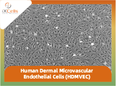
Human Dermal Microvascular Endothelial Cells (HDMVEC) from blood vessels of skin, form the interface between intravascular and extravascular compartments in skins. Compared to endothelial cells elsewhere in the body, HDMVEC exhibit several skin specific characteristics. They actively participate in a variety of physiological processes including wound healing, control of hemostasis, temperature regulation, and modulation of inflammation/leukocyte trafficking [1]. Via proliferation, quiescence, apoptosis, and senescence, HDMVEC show remarkable phenotypic and functional heterogeneity, which in turn allows the cutaneous microvasculature to be in a dynamic balance between maintenance and remodeling [2,3].
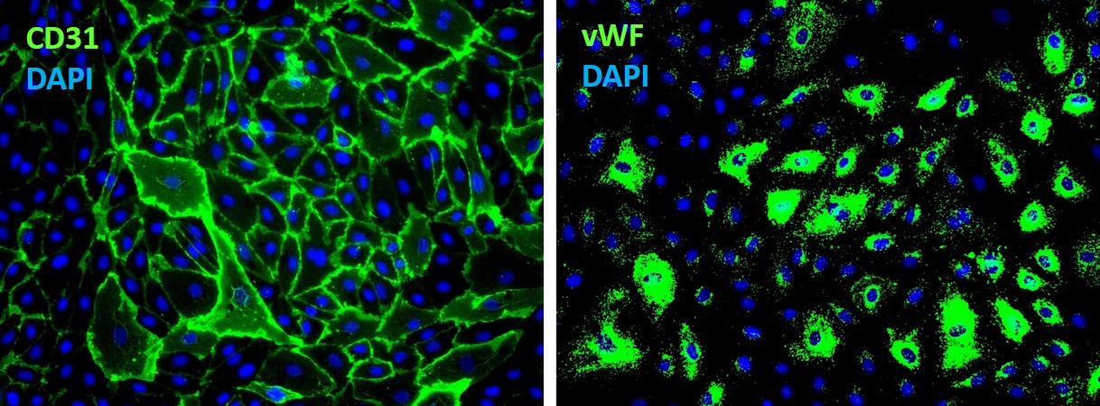
iXCells Biotechnologies provides high quality HDMVEC isolated from human skin blood vessels and cryopreserved at P2, with ≥0.5 million cells in each vial. These HBMVEC express von Willebrand Factor (vWF), CD31 (PECAM), and E-Cadherin. They are negative for HIV-1, HBV, HCV, mycoplasma, bacteria, yeast, and fungi and can be further expanded for no more than 3 passages in Endothelial Cell Growth Media under the condition suggested by iXCells Biotechnologies. Further expansion may decrease the purity.

Figure 1. (A) Phase contrast image of HDMVEC on day 1 and day 2 post recovery. (B) Immunofluorescence staining of HDMVEC with antibodies against VE-Cadherin (Green).
Product Details
| Tissue | Human skin blood vessels |
| Package Size | 0.5 millioncells/vial |
| Passage Number | P2 |
| Shipped | Cryopreserved |
| Storage | Liquid nitrogen |
| Growth Properties | Adherent |
| Media | Endothelial Cell Growth Media |
References
[1] Avril M, Tripathi AK, Brazier AJ, Andisi C, Janes JH, Soma VL, Sullivan DJ Jr, Bull PC, Stins MF, Smith JD. (2012) “A restricted subset of var genes mediates adherence of Plasmodium falciparum-infected erythrocytes to brain endothelial cells.” Proc Natl Acad Sci USA. 109: E1782-90.
[2] Claessens A, Adams Y, Ghumra A, Lindergard G, Buchan CC, Andisi C, Bull PC, Mok S, Gupta AP, Wang CW, Turner L, Arman M, Raza A, Bozdech Z, Rowe JA. (2012) “A subset of group A-like var genes encodes the malaria parasite ligands for binding to human brain endothelial cells.” Proc Natl Acad Sci USA. 109: E1772-81.
[3] Laranjeira MS, Fernandes MH, Monteiro FJ. (2012) “Reciprocal induction of human dermal microvascular endothelial cells and humanmesenchymal stem cells: time-dependent profile in a co-culture system.” Cell Prolif. 45: 320-34.


Reviews
There are no reviews yet.