
10HU-007
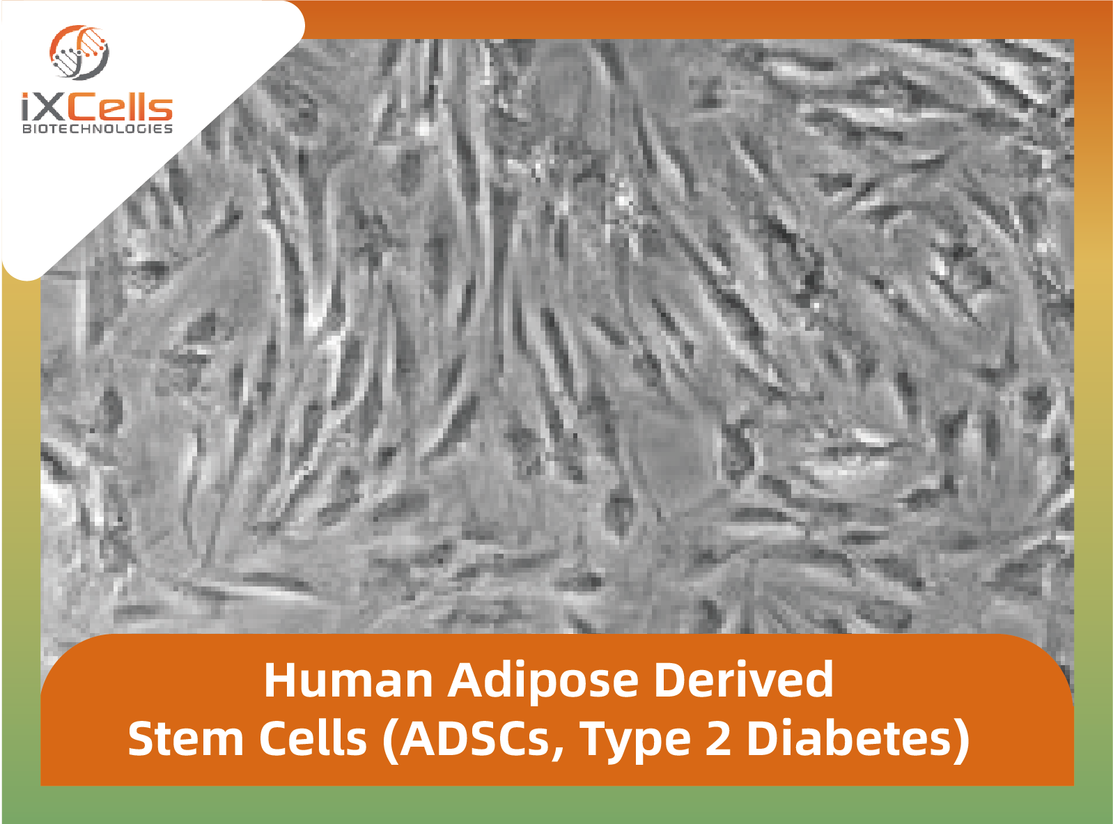
Human Adipose Derived Stem Cells (ADSCs, Type 2 Diabetes)
Cryopreserved, 0.5 million cells/vial
产品与技术资料CELL CULTURE / PRODUCT
01 / PRODUCT INFORMATION
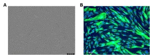 Figure 1. Human Pancreatic Microvascular Endothelial Cells (HPaMEC). (A) Phase contrast image of HPaMEC. (B & C) Immunofluorescence staining with antibodies against VE-Cadherin (B) and vWF/Factor VIII (C).
| Tissue | Human pancreatic tissue |
| Package Size | 5 x 10^6 cells/vial |
| Passage Number | P2 |
| Shipped | Cryopreserved |
| Storage | Liquid Nitrogen |
| Growth Properties | Adherent |
| Media | Endothelial Cell Growth Medium (Cat# MD-0010) |
02 / PRODUCT INFORMATION
Product Overview
The pancreas functions as both an endocrine gland and a digestive organ, secreting hormones and producing pancreatic juice to aid in digestion.[1,2] The pancreatic islets, which serve as the endocrine component of the pancreas, produce essential hormones such as glucagon, insulin, and amylin [1,2]. Human Pancreatic Microvascular Endothelial Cells (HPaMECs) play a critical role in supporting the islets by delivering oxygen and nutrients to pancreatic tissue, influencing beta cell function and proliferation, and regulating insulin gene expression during islet development. Additionally, HPaMECs produce various growth factors, optimize blood glucose sensing, and help regulate blood glucose levels. HPaMECs can serve as an in vitro model for studying islet biology, pancreatic cancer, transplantation, and regenerative medicine.
iXCells Biotechnologies provides high quality HPaMECs, which are isolated from human pancreas and cryopreserved at P2, with ≥ 0.5 million cells in each vial. These HPaMECs express von Willebrand Factor (vWF), VE-Cadherin and CD31/PECAM-1. They are negative for HIV-1, HBV, HCV, mycoplasma, bacteria, yeast, and fungi, and can be further expanded for no more than 3 passages under the conditions suggested by iXCells Biotechnologies. Further expansion may decrease the purity.
Figure 1.Phase contrast images of Human Pancreatic Microvascular Endothelial Cells (HPaMECs). The cells were recovered and seeded at 10,000 cells/cm2 following iXCells’ protocol. The images were taken at the indicated time post recovery.
Figure 2.Immunofluorescence staining of HPaMECs using antibodies against VE-Cadherin (green), vWF (green) and CD31/PECAM-1 (green) with nuclei counterstained by DAPI (blue).
Product Details
| Organism | Homo Sapiens, Human |
| Cell Type | Endothelial Cells |
| Tissue | Human Pancreas |
| Disease | Normal |
| Package Size | 0.5 x 10⁶ cells/vial |
| Passage Number | P2 |
| Growth Properties | Adherent |
| Product Format/Shipped | Cryopreserved |
| Storage | Liquid Nitrogen |
References
[1] Zanone M, Favaro E, Camussi G (2008). “From endothelial to beta cells: insights into pancreatic islet microendothelium.” Curr Diabetes Rev. 4(1): 1-9.
[2] Konstantinova I and Lammert E. (2004). “Microvascular development: learning from pancreatic islets.” Bioessays, 26(10):1069-1075.
03 / PRODUCT INFORMATION
04 / PRODUCT INFORMATION
此 Bioz 组件为宽版,可左右滑动查看完整内容。
Bioz 实时文献组件 · 可在组件内筛选和滚动浏览;无法载入时可在 Bioz 打开。
05 / PRODUCT INFORMATION
SAME COLLECTION
10HU-007
Cryopreserved, 0.5 million cells/vial
产品与技术资料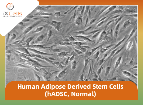
10HU-001
Human Adipose Derived Stem Cells (hADSC, Normal)
Cryopreserved, 0.5 million cells/vial · 2 种现有规格
产品与技术资料
10HU-020
Human Aorta Endothelial Cells
Cryopreserved, 0.5 million cells/vial
产品与技术资料
10HU-190
Human Aortic Smooth Muscle Cells (HASMC)
Cryopreserved, 0.5 million cells/vial
产品与技术资料BIOMO / 实验需求

